The number of viable bacteria is one of the important attributes of probiotic products, because the number of viable bacteria may affect the effect of probiotic products. The number of viable bacteria is of great significance not only to probiotic companies and consumers, but also to regulatory authorities. So how to detect the number of viable bacteria? Are there any other methods besides plate culture?
Today, we are jointly concerned about the detection of viable cell counts, and hope that this article can bring some inspiration and help to related industry professionals and readers.
① Live bacteria detection
At present, the definition of probiotics recognized by the probiotic industry was proposed by the United Nations Food and Agriculture Organization/World Health Organization joint working group in 2001: Probiotics refer to active microorganisms that produce health benefits to the host when ingested in sufficient quantities [ 1]. Therefore, by definition, “activity” is an important characteristic of probiotics.
In addition, Article 12 of the “Probiotics Health Food Application and Review Regulations (Trial)” issued by my country in 2005 also clearly stipulates [2] that the number of live bacteria of probiotic health foods shall not be allowed during the shelf life. Less than 10[6] CFU/mL(g).
Therefore, the accurate determination of the number of viable bacteria in probiotic products is of great significance to manufacturers and consumers, as well as regulatory authorities.
However, with the continuous development of technology, some new methods for detecting the number of live bacteria have emerged, some of which have certain application prospects and have the opportunity to be widely used in the detection of live bacteria in probiotic products in the future. Today, we will introduce several methods for the detection of live bacteria and discuss the advantages and disadvantages of different methods.
② Based on separation culture: plate count
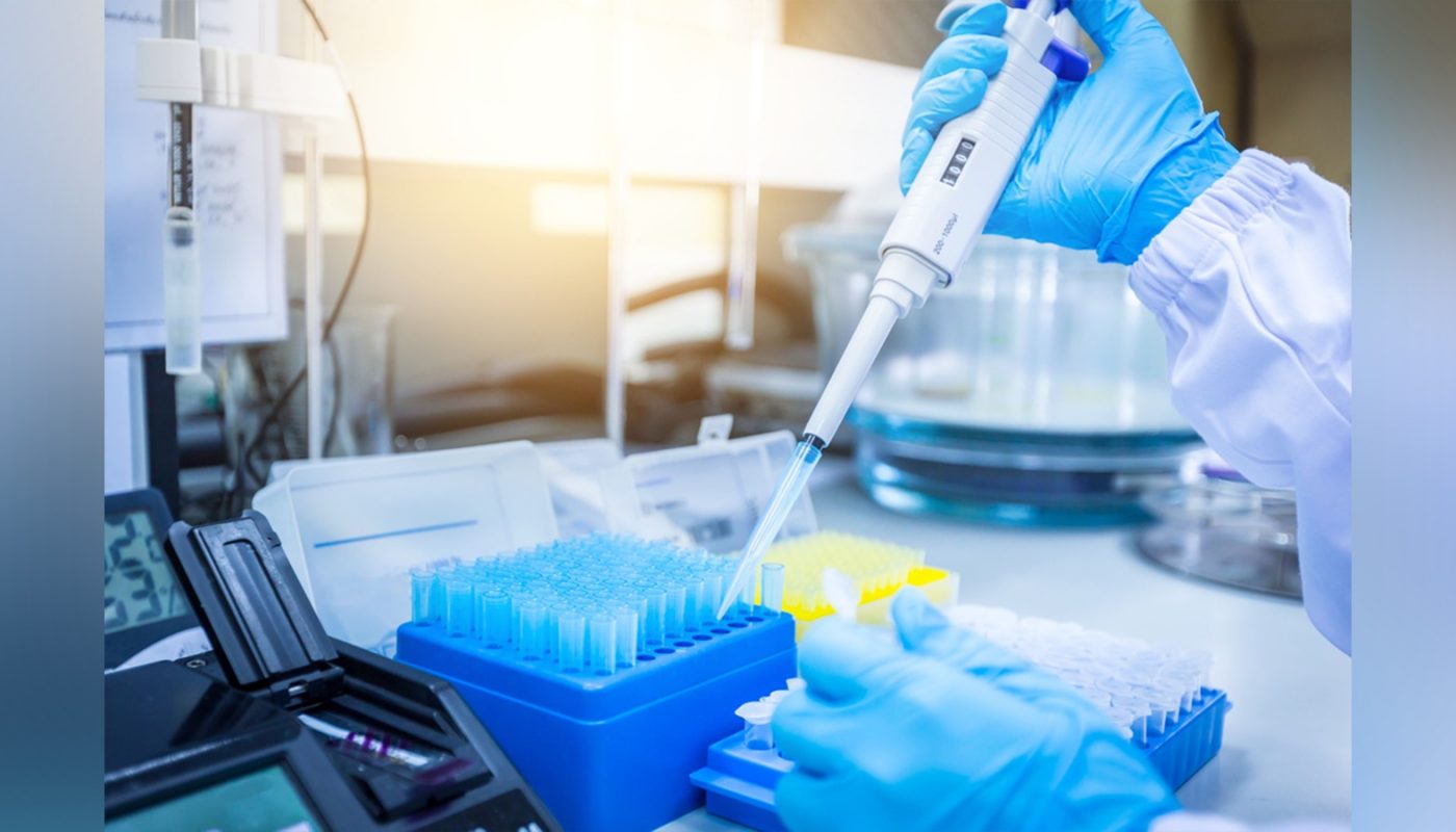
The plate colony counting method is to apply a certain amount of diluted sample solution to the plate for culture after appropriate serial dilution of the sample, and count the colony forming units (CFU) to obtain the estimated value of the number of live bacteria, and Expressed as a method of colony forming units per gram or milliliter of the original sample.
The key to this method is the proper dilution factor of the original sample-it is advisable to control the number of bacterial colonies after culture to 30-300.
This live bacteria detection method is currently one of the most widely used and classic microbiological detection techniques. In addition to being used to detect the number of live bacteria in food, it is often used for water quality detection [4]. This method has great advantages of low cost and easy operation, but it also has certain limitations.
The plate colony counting method has the disadvantages of time-consuming and difficult to cope with slow-growing and viable “non-cultivable” microorganisms. Not only that, with the continuous deepening of research on probiotics, more and more diverse microbial strains (strains) will appear in the future. For these uncultured strains, experimental conditions need to be explored and tried.
In addition, the result of this method is the total number of live bacteria, which cannot accurately distinguish the number of live bacteria of different strains of bacteria in probiotic products. At present, more and more probiotic products use multiple strains, so other live bacteria are urgently needed. Detection methods to make up for the above-mentioned shortcomings.
③ Based on gene expression activity: qPCR
The transcriptome can reflect the viability of microorganisms. Therefore, some researchers are currently trying to use bacterial messenger RNA (mRNA) to determine the number of viable bacteria.
Quantitative reverse transcription PCR (RT-qPCR) is a common research method used to detect gene transcription. In this method, messenger RNA (mRNA) is first transcribed into complementary DNA (cDNA) by reverse transcriptase. Subsequently, the cDNA was used as a template for the qPCR reaction. Therefore, we can select the mRNA of the specific gene of the microorganism as the detection target, and use this method to determine the number of viable bacteria in the sample.
In order to improve the accuracy of mRNA-based detection methods, RT-qPCR can be combined with technologies such as propidium azide iodide staining (PMA, which can selectively label dead cells) and 16S rRNA sequencing [5] to set a dead bacteria positive control group , To distinguish live bacteria.
At present, some documents have adopted this method to detect the number of viable bacteria in products, such as Enterococcus faecalis, which is widely used in livestock and poultry breeding [6].
This detection method based on gene expression activity has the advantages of high sensitivity, strong specificity, and rapidity; however, small changes in the sample preparation process will affect the final measurement results, and require high experimental environment and operators. At the same time, different detection targets and corresponding standards need to be formulated according to different microorganisms.
④ Flow cytometry sorting technology
With the development of technology, flow cytometry sorting technology has become one of the common experimental methods in the field of life sciences. On the basis of staining or fluorescent labeling, flow cytometry sorting technology can be used to count bacteria.
Flow cytometric sorting technology is a kind of single cell or particle dyed by fluorescent pigment under high-energy laser irradiating high-speed flow state, and measuring the intensity of scattered light and fluorescence emitted by it, so as to improve the physical performance of the cell (or particle). Modern cell analysis technology for qualitative or quantitative detection of physiological, biochemical, immune, genetic, molecular biological traits and functional status. It can separate the subgroups of luminescent particles according to the fluorescence intensity and wavelength of the emitted light, and can achieve monoclonal analysis. Select to classify, quantify, and separate cells in complex samples.
The flow cytometry sorting technology has the advantages of high accuracy and fast speed, allowing the detection of a large number of bacteria at one time, sorting different bacteria, and detecting the advantages of different physiological activities and bacterial sizes of live bacteria [7].
However, due to the complicated and expensive flow cytometry sorting technology, it is currently mainly used in scientific research and has not yet been widely used for viable cell count testing of probiotic products. However, there is a precedent in foreign countries that uses this as a factory testing standard.
⑤ Raman spectroscopy detection method
In 1928, when studying the passage of monochromatic light through a transparent liquid medium, the Indian physicist CV Raman discovered for the first time that a small part of the photons in the incident light produced an inelastic collision with the medium, that is, when the incident light irradiated on the medium, it collided with the medium. The molecules or atoms of the medium exchange energy, and the vibration frequency of the scattered light is different from that of the incident light-this is the famous Raman spectrum effect.
Based on this theory, the developed analysis technology is called Raman spectroscopy technology, which can be widely used in the detection of liquids, gases and solids, and can obtain molecular fingerprint information with a vibration frequency of 50-4000cm[-1][ 8]. Therefore, it has been widely used in the identification and research of chemical and biological molecules in many fields, such as food testing, biomedicine, environmental sanitation and petrochemicals.
Raman spectroscopy can also be used to count live bacteria. Some researchers have used the difference between the surface charge of live bacteria and dead bacteria to reduce the surface of the live bacteria after adsorbing silver ions, and test the Raman enhancement signal. The results show that the completely inactivated bacteria have almost no Raman enhancement signal. The Raman enhancement signal on the surface of live bacteria is very abundant, which can be used to distinguish live bacteria [9].
Some researchers combine separation culture with Raman spectroscopy technology, culture Salmonella with nutrient broth prepared from heavy water, label it with deuterium, and combine it with confocal Raman spectroscopy to characterize Raman spectrum peaks with deuterium. Surveillance distinguishes Salmonella as dead or live [10].
This technology can accurately reflect the differences in the chemical composition and molecular structure of samples at the molecular level, and the emergence of surface-enhanced Raman spectroscopy and confocal micro-Raman spectroscopy makes the process of analysis and identification more intuitive and Easy to observe and control, and the results are more significant, these advantages have greatly promoted the development of Raman spectroscopy technology.
However, as the technology is still under study, there are still some problems to be overcome. For example, this technology is difficult to distinguish the superposition of Raman peaks of different components, and it is easy to cover up the peaks of certain substances to be tested, resulting in the incomplete display of certain component information, which affects the accuracy of the final result.
At present, many researchers are actively overcoming these limitations and promoting the application of Raman spectroscopy technology, and believe that this method is expected to become a new generation of viable cell count detection method instead of culture method.
⑥ Other methods: products of bacteria
In addition to Raman spectroscopy, there are some new methods for detecting live bacteria. Probiotics may secrete some substances or emit some gases during the growth and fermentation process. Based on these biomarkers, the number of live bacteria can also be determined indirectly by detecting the amount of secretions or gases produced by the live bacteria.
Potential secretions that can be used for live bacteria detection are: Type III secreted protein SpaO, protein precursor translocation enzyme.
In addition, some probiotics will produce short-chain fatty acids and other acidic substances, which will reduce the pH of the sample. Use bromocresol purple, phenolphthalein, nitrophenol, etc. as pH indicators, or use a high-precision pH meter to detect the pH value. The amount of change, thereby converting the amount of acid production, and extrapolating the number of viable bacteria.
This type of new detection method has a huge imagination, but it is still very immature, and the standard for distinguishing dead and living bacteria using biomarkers is still not comprehensive. However, it is believed that in the near future, these live bacteria detection methods will gradually mature, providing us with more possibilities for detecting the number of live bacteria.


